Affiliated Foot Care Center
Call 860•349•8500 or 203•294•4977

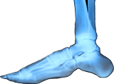
The ankle, or the talocrural region is where the foot and the leg meet. The Ankle contains three joints, the ankle joint proper or the talocrural joint, the subtalar joint, and the inferior tibiofibular joint. The movements produced at this joint are dorsiflexion and plantarflexion of the foot.
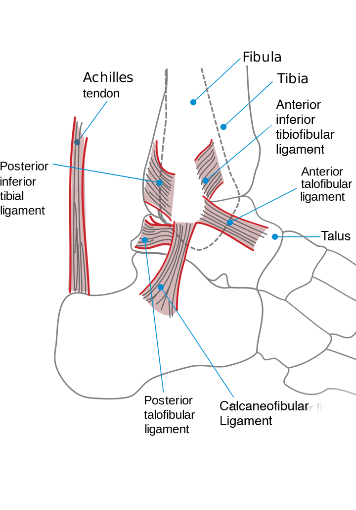
The bony architecture of the ankle consists of three bones: the tibia, the fibula, and the talus. The articular surface of the tibia is referred to as the plafond. The medial malleolus is a bony process extending distally off the medial tibia. The distal-most aspect of the fibula is called the lateral malleolus. Together, the malleoli, along with their supporting ligaments, stabilize the talus underneath the tibia.
The bony arch formed by the tibial plafond and the two malleoli is referred to as the ankle "mortise" (or talar mortise). The mortise is a rectangular socket. The ankle is composed of three joints: the talocrural joint (also called tibiotalar joint, talar mortise, talar joint), the subtalar joint (also called talocalcaneal), and the Inferior tibiofibular joint.
All of the bones in your ankle are covered with articular cartilage.
A sprained ankle is one of the most common orthopedic injuries. Every day, about 25,000 people in the U.S. suffer an ankle sprain. Ankle sprains occur in both athletes and those with sedentary lifestyles, and they can occur during sports or when walking to carry out daily activities.
A sprain is actually an injury to the ligaments of the ankle joint, which are elastic, band-like structures that hold the bones of the ankle joint together and prevent excess turning and twisting of the joint. In normal movement, the ligaments can stretch slightly and then retract back to their normal shape and size. A sprain results when the ligaments of the ankle have been stretched beyond their limits. In severe sprains, the ligaments may be partially or completely torn.
Depending on the severity of the break and how many bones have been broken, fractures can be treated either surgically or nonsurgically. Doctor Fosdick may treat the break without surgery by immobilizing the ankle if only one bone is broken, and if the bones are not out of place and the ankle is stable. This is done by the use of a brace that works as a splint or by using a cast to immobilize the joint. If your ankle is unstable, Dr. Fosdick can use surgery to further stabilize the bones of your foot with screws and plates and will then either use a cast or a brace for after surgery.
It usually takes at least six weeks for the bones to heal. Dr. Fosdick will probably ask you to keep weight off the ankle during that time so the bones can heal in the proper alignment. Ligaments and tendons can take longer to heal after a fracture is fully mended. It can take as long as two years to completely recover full pain-free motion and strength after an ankle fracture, although most people are able to resume their normal daily routine within three to four months. While you are recovering and after the bones have knit, Dr. Fosdick may perscribe physical therapy to assist in your recovery.
To diagnose a sprained ankle, Dr Fosdick may take X-rays or other imaging studies to confirm that a bone has not been broken.
Ice can help reducing swelling. Cycles of 10-15 minutes on and 10-15 minutes off are recommended. Icing an ankle too long may cause cold injuries.
An ankle brace can be very helpful for the treatment and prevention of a sprained ankle injury. Walking is inadvisable, as it may cause the ankle sprain to become worse - with increased pain, and possible further damage to the compromised tissues. Braces and crutches give the leg exercise, yet keep the damaged part from moving and becoming further injured.
Dr Fosdick has found that ankle sprains heal very quickly with high-powered laser therapy.
Regular checkups after you hae healed are advisable to prevent a reinjury.